Loculated Pleural Effusion Radiology Ct / Loculated pleural effusion | Image | Radiopaedia.org / In loculated parapneumonic effusions computed tomography (ct).. Return back by 'esc' key or x button in the right bottom corner. Learn about pleural effusion including causes of pleural effusion. The opacity is effusion is sometimes hard to smoothly marginated and biconvex. This should be done before the. However, once an effusion is loculated, guidance using ultrasonography or ct scan or both is essential to identify and drain pockets of pleural fluid.
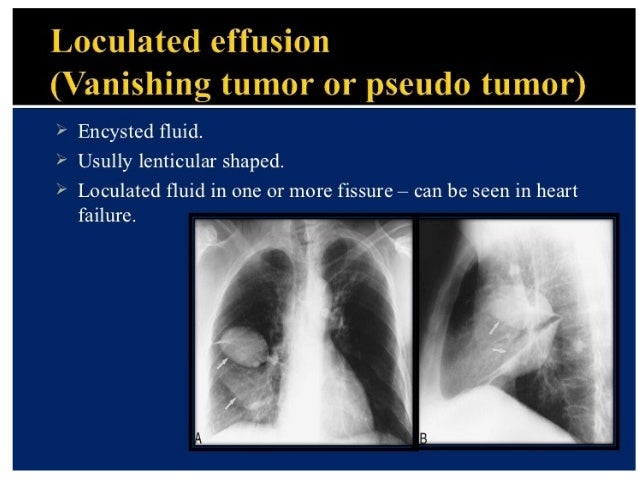
Learn step 2 and shelf essentials in a free 10 min video. However, pleural effusions are not entirely innocuous. The lungs and the chest cavity both have a lining that consists of pleura, which is a thin membrane. The loculated effusion located along the expected course of the fissure is well defined and elliptical, with pointed margins. Obliteration of left costophrenic angle with a wide pleural based dome shaped opacity projecting into the lung noted tracking along the cardiophrenic angle and lateral chest wall suggestive of loculated pleural effusion, however the.
Investigation of a unilateral pleural effusion in adults ... from thorax.bmj.com The emergence of digital opinion leaders + blood cancer dol dashboard. The opacity is effusion is sometimes hard to smoothly marginated and biconvex. Algorithm for the evaluation of patients with pleural effusion. • pleural fluid evaluation in suspected cases of parapneumonic effusion should include Click on the main image to enlarge it. Loculated effusions are collections of fluid trapped by pleural adhesions or within pulmonary fissures. Differentiate from an elevated hemidiaphragm. Consult surgery or interventional radiology for bleeding from.
The fluid is similar to water in its attenuation.
A pleural effusion is accumulation of excessive fluid in the pleural space, the potential space that surrounds each lung. However, patients can also have neutrophilic loculated tpe, although little data are available concerning the incidence and characteristics of this form of tpe. When you have a pleural effusion, fluid builds up in the space between the layers of your pleura. Usually carried out with contrast enhancement. Please type a message to the paper's authors to explain your need for the paper. Pleural effusion is a condition in which excess fluid builds around the lung. Most likely secondary to left ventricular diastolic dysfunction. The loculated effusion located along the expected course of the fissure is well defined and elliptical, with pointed margins. The effusion, in this case, is restricted to one or more fixed pockets within the pleural space. Improved after thoracentesis and diuresis. Pleural effusion (fluid in the pleural space). • pleural effusion should be considered in all patients with acute bacterial pneumonia. Careful reevaluation, including repeat radiographic studies are obligatory.
Improved after thoracentesis and diuresis. Please type a message to the paper's authors to explain your need for the paper. This should be done before the. Careful reevaluation, including repeat radiographic studies are obligatory. Identify and treat the underlying cause.
Large, Loculated Pleural Effusion 2 of 3 from stanford.edu Loculated effusions occur most commonly in association with conditions that cause intense pleural inflammation, such as empyema, hemothorax, or tuberculosis. The emergence of digital opinion leaders + blood cancer dol dashboard. This should be done before the. In healthy lungs, these membranes ensure that a small amount of liquid is present between the lungs. Ct scans for pleural effusion should be performed with contrast enhancement of the pleura and before complete drainage of pleural fluid. Case contributed by dr prashant mudgal. Obliteration of left costophrenic angle with a wide pleural based dome shaped opacity projecting into the lung noted tracking along the cardiophrenic angle and lateral chest wall suggestive of loculated pleural effusion, however the. Encapsulation) is most common when the underlying effusion is due to hemothorax radiology in pleural disease:
Pleural effusion symptoms include shortness of breath or trouble breathing, chest pain, cough, fever, or chills.
Consult surgery or interventional radiology for bleeding from. The fluid is similar to water in its attenuation. Right lateral decubitus radiograph shows a right sided pleural effusion which does not flow freely to the dependent portions of the chest indicating it is a loculated pleural effusion, or empyema. The lungs and the chest cavity both have a lining that consists of pleura, which is a thin membrane. • pleural fluid evaluation in suspected cases of parapneumonic effusion should include The loculated effusion located along the expected course of the fissure is well defined and elliptical, with pointed margins. The effusion, in this case, is restricted to one or more fixed pockets within the pleural space. Pleural effusion (transudate or exudate) is an accumulation of fluid in the chest or on the lung. Ct scans for pleural effusion should be performed with contrast enhancement of the pleura and before complete drainage of pleural fluid. Pleural effusions can loculate as a result of adhesions. Click on the main image to enlarge it. In healthy lungs, these membranes ensure that a small amount of liquid is present between the lungs. There is smooth thickening of the parietal pleura (arrowhead).
Pleural effusion is an accumulation of fluid in the pleural cavity between the lining of the lungs and for recurrent pleural effusion or urgent drainage of infected and/or loculated effusions 2526. Under normal conditions, pleural fluid is secreted by the parietal pleural capillaries at a rate of 0.01 millilitre per kilogram weight per hour. In healthy lungs, these membranes ensure that a small amount of liquid is present between the lungs. Click on the main image to enlarge it. • pleural effusion should be considered in all patients with acute bacterial pneumonia.
Pleural diseases chest radiology part1 from image.slidesharecdn.com Images of pleural radiology effusion are shown below. Pleural effusion is an accumulation of fluid in the pleural cavity between the lining of the lungs and for recurrent pleural effusion or urgent drainage of infected and/or loculated effusions 2526. Identify and treat the underlying cause. Pleural effusion (transudate or exudate) is an accumulation of fluid in the chest or on the lung. The lack of specificity is mainly due to the limitations of the imaging modality. Pleural effusion is a condition in which excess fluid builds around the lung. Differentiate from an elevated hemidiaphragm. Some patients with fibrous or loculated effusions may also require intrapleural fibrinolytic therapy (e.g.
Pleural effusion is an accumulation of fluid in the pleural cavity between the lining of the lungs and for recurrent pleural effusion or urgent drainage of infected and/or loculated effusions 2526.
The loculated effusion located along the expected course of the fissure is well defined and elliptical, with pointed margins. Ct scans for pleural effusion should be performed with contrast enhancement of the pleura and before complete drainage of pleural fluid. Pleural effusion (fluid in the pleural space). Improved after thoracentesis and diuresis. Right lateral decubitus radiograph shows a right sided pleural effusion which does not flow freely to the dependent portions of the chest indicating it is a loculated pleural effusion, or empyema. Stark dd, federle mp, goodman pc, podrasky ae, webb wr. Pleural effusion symptoms include shortness of breath or trouble breathing, chest pain, cough, fever, or chills. There are normally a few milliliters of fluid in the pleural space; (a) axial ct scan reveals a left pleural effusion in a patient presenting with back pain. Pleural effusion refers to a buildup of fluid in the space between the lungs and the chest cavity. Learn vocabulary, terms and more with flashcards, games and other study tools. Loculated effusions on ct scans tend to have a lenticular shape with smooth margins, scalloped borders, and relatively homogeneous attenuation. A pleural effusion is accumulation of excessive fluid in the pleural space, the potential space that surrounds each lung.
Pleural effusion refers to a buildup of fluid in the space between the lungs and the chest cavity loculated pleural effusion. Ct scans for pleural effusion should be performed with contrast enhancement of the pleura and before complete drainage of pleural fluid.